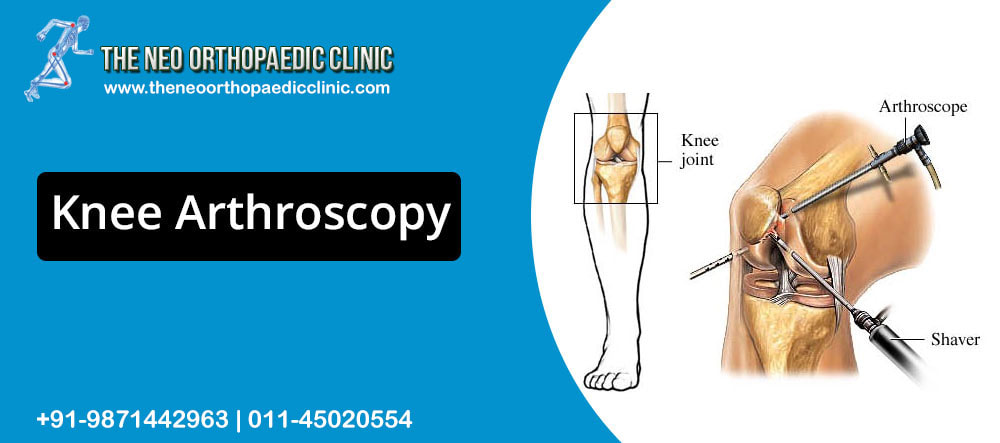
 Arthroscopy in Delhi is a surgical intervention allowing the exploration of the joint using a tube a few millimeters in diameter, introduced into the knee through several tiny holes (2 to 4) (about 1 cm). This tube, fitted with optics coupled to a miniaturized video camera, is connected to a color television screen on which the main elements of the knee will be viewed:
ARTHROSCOPY THEREFORE ALLOWS:
THESE INTERVENTIONS CAN BE:
IN PRACTICE Arthroscopy in West Delhi is performed in the operating room, under anesthesia. The terms of this will be decided between you and the anesthesiologist. You will be admitted to the hospital the same morning; you must bring the x-rays as well as the biological examinations in your possession and report the usual treatments in progress. On the day of the arthroscopic surgery in Delhi you must be on an empty stomach and respect the instructions of orthopaedic surgeon in Delhi. As soon as you get back to your room, you can start to gently mobilize your knee, walking being allowed with the help of the nurse or physiotherapist a few hours later. The exit from the hospital will generally be rapid. During this, you will be given:
During the first week, you will be advised:
Arthroscopy is a surgical procedure. If the hazards inherent in this technique are rare, do not hesitate to report any local incident to orthopaedic in Delhi that would worry you: persistent fever, increasing pain, hematoma or significant swelling of the knee (moderate, painless swelling is however usual with possible sensation of ” splashing “due to temporary persistence of fluid in the knee).
0 Comments
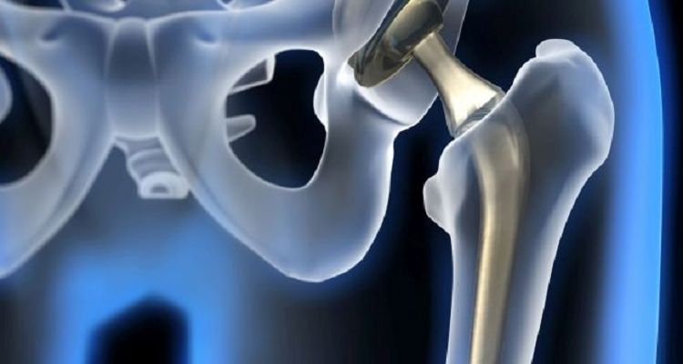 Pain in the hip joint is most often the result of osteoarthritis and can seriously affect your ability to lead a full and active lifestyle. Hip osteoarthritis is called coxarthrosis in medicine. Endoprosthesis of the hip joint can help relieve pain and return to normal life. Over the past 20 years, thanks to the introduction of new materials and techniques into practice, the results of endoprosthetics operations have significantly improved. Endoprosthesis of the hip joint is becoming more and more prevalent as the world's population is aging. At the moment, hip replacement surgery in Delhi is the most commonly performed in the world. HIP ANATOMY The hip joint is spherical in structure, so movements in it are possible in many planes. The joint is formed by the acetabulum, forming, as it were, a deep bowl and the head of the femur, which has the shape of a ball, explains the orthopaedic in Delhi. The femoral head is connected to its main part (diaphysis) using a short portion of the bone called the femoral neck. Strong and thick muscles and tendons surround the joint. The surfaces of the acetabulum and femoral head are covered with articular cartilage. The thickness of the articular cartilage is about half a centimeter in large joints. The articular cartilage is a hard and smooth material covering the bones in the joint area. The articular cartilage allows the bones coated with it to glide smoothly relative to each other without being damaged. The color of the articular cartilage is white and shiny. The joint is surrounded by a dense waterproof capsule, inside which a special fluid is produced that lubricates the mating surfaces. The bones in the joint hold tight ligaments and muscles together. The design of the hip joint provides extremely high mobility while maintaining satisfactory stability. The powerful muscles around the joint allow us to move for a long time in an upright position, and also, if necessary, accelerate when running and jumping. Important nerves and blood vessels also pass around the joint. WHEN CAN ENDOPROSTHETICS REQUIRED? The main indications for hip replacement in Delhi are arthrosis of the hip joint (coxarthrosis), fracture of the femoral neck, aseptic necrosis of the femoral head. With arthrosis, degenerative changes in the articular cartilage occur, which ultimately leads to cartilage wear. Bone growths (osteophytes) form around the joint. Due to the deterioration of the cartilage, a decrease in its thickness, a significant decrease in smoothness, as well as a change in the shape of the articular surfaces, the friction in the joint increases, which leads to pain and a progressive violation of the movements in the joint. Aseptic necrosis of the femoral head is another cause of destruction of the hip joint. With this disease, the femoral head loses blood supply and actually collapses. The shape of the femoral head changes, the bone tissue making up the head is resorbed. The articular surfaces of the acetabulum and the femoral head cease to correspond in shape, pain and impaired movement in the joint appear. The causes of the disease can be previous hip dislocations, birth injuries, prolonged treatment with corticosteroids, as well as some infections. The main goal of replacing the joint in any of the degenerative diseases with an artificial one is to reduce pain and return movements. To do this, damaged surfaces are replaced with artificial ones, as a result of which the smoothness and painlessness of movements in the joint returns. Fracture of the femoral neck is also an indication for joint replacement surgery in Delhi. In case of fractures of the femoral neck, the blood supply to the head is disturbed, in connection with which its gradual destruction occurs. Fracture fusion in these conditions is impossible, hip replacement surgery in Delhi is the only way to activate the patient and return him to everyday activity. PREPARATION FOR HIP JOINT REPLACEMENT The decision about the operation is made by the orthopaedic doctor in Delhi together with the patient. After clarifying the medical history, the doctor performs a thorough clinical examination to measure the current range of motion, the level of pain, the patient’s functionality. During the examination of the patient, the orthopaedic surgeon in Delhi examines the radiographs, as well as the data of CT and MRI studies. A thorough and complete medical examination before surgery is also required. This is done in order to minimize the risk of complications during hip replacement surgery in Delhi. If a long-term operation or hemoglobin level of the patient is expected to be below normal values, a blood transfusion may be required after or during the operation. Mandatory prophylaxis of thromboembolic complications. TYPES OF ENDOPROSTHESES There are several main types of endoprostheses - cementless and cement. Cemented endoprostheses are held in the bone using a special cement that fixes the metal to the bone. The surface of cementless prostheses is made in such a way that the bone tissue grows into it over time, due to which the prosthesis is held in the bone. In order for the endoprosthesis to grow, the bone is treated with special tools. Both types of fixation of endoprostheses are widely used in medical practice. Also, in some cases, a combination may be used where, for example, the acetabular component (cup) is fixed with cement, and the femoral component (leg) is cementless. The decision about whether to use a cement or cementless endoprosthesis is made by the orthopaedic surgeon in Delhi based on the age, lifestyle of the patient and the quality of his bones. The endoprosthesis consists of two main parts. The acetabular component (cup) replaces the articular surface of the acetabulum. The shell of the acetabular component is made of metal, inside of which is placed a plastic or ceramic insert that is directly in contact with the femoral component. The femoral component replaces the head and neck of the femur, usually made entirely of metal. In some designs of the endoprosthesis, the head may be made of ceramic. Endoprosthetics can be total when both components are replaced, and unipolar. With unipolar endoprosthetics (hemiarthroplasty), only the femoral component changes. Hemiarthroplasty is usually performed for fractures of the femoral neck in elderly and debilitated patients. With this type of endoprosthetics, the earliest verticalization of the patient is allowed, the very next day. This significantly reduces the risk of thromboembolic and hypostatic complications in elderly debilitated patients with femoral neck fractures. Equally important is the shorter operation time for hemiarthroplasty compared with total arthroplasty, which also reduces the risks during anesthesia and blood loss during surgery. Currently, our clinic uses modern cemented bipolar hip arthroplasty. A bipolar endoprosthesis is a modern type of unipolar prosthesis in which the head is double. A similar design of the endoprosthesis increases the life of the prosthesis, increases its stability and range of motion. MORE ABOUT HIP JOINT OPERATION The orthopaedic surgeon in Delhi performs access to the hip joint, a skin incision is performed in the upper third of the thigh. After the hip joint is exposed, surgeons dislocate the worn femoral head from the acetabulum. Then, a resection of the damaged head and neck of the femur is performed with a special electric saw. Then, using special mills, the acetabulum is processed. During the treatment, the worn cartilage is completely removed and a hemisphere is formed into which the acetabular component will be implanted. After the formation of the acetabulum, the surgeon fills the cavity with bone cement and establishes a suitable acetabular component. At this stage, the correct spatial orientation of the acetabular component at the right angle is important. This affects the life of the endoprosthesis and the likelihood of complications in the postoperative period. After cement hardens and fixation of the acetabular component, the surgeon proceeds to the femur. At this stage, the bone canal of the femoral canal is developed with special rasps to the required size. Next, cement is placed in the prepared canal in the femur and the femoral component is installed. A head of the required size is selected and the femoral component is set into the acetabular. After the orthopaedic surgeon in West Delhi checks the stability of the thigh and range of motion. As soon as the surgeon is convinced that everything is set properly, the wound is sutured in layers. Drainages are established for a day. The patient is sent to a special ward in the postoperative ward, explains the orthopaedic in Delhi. From the first day, rehabilitation of the patient begins. What is knee arthroscopy? Knee arthroscopy in Delhi is a minimally invasive procedure that allows access and treatment of injuries that affect the various structures of the joint. For this, 2 or 3 small incisions of less than one centimeter are made that allow access to the joint. Most knee surgeries that do not involve the knee replacement in Delhi are performed with a complete or partial approach with knee arthroscopy. It is the technique of choice to deal with many injuries because it allows a better and greater visualization of the joint. In a non-aggressive way, all the points of the knee can be accessed to make a cartilaginous cleaning, small perforations of the bone that has lack of cartilage (microfractures), stabilize the cartilage and apply substances or elements that regenerate cartilage (plasma rich in platelets or stem cells). Likewise, knee arthroscopy in Delhi is also used to support other open techniques, since it improves the diagnosis and prognosis of the patient’s injury, as it is less aggressive. Surgery can be performed under local, regional or general anesthesia, depending on the injury and the patient himself. The anesthesiologist will decide the best method for the patient, provided he suffers as little as possible, explains the best knee surgeon in Delhi. Why is it done? Knee arthroscopy in Delhi is used to resolve knee injuries. Thus, meniscus injuries are one of the most common pathologies and, thanks to arthroscopy, it is possible to preserve most of the menisci, since the resection is not complete but partial. Meniscal sutures and the possibility of transplanting the meniscus with knee arthroscopy are common techniques that allow better protection of the cartilage of the joint. Another of the most dangerous injuries related to sports is the rupture of the anterior cruciate ligament. If this is injured, it causes instability in the knee that makes it impossible for the patient to practice practically any sport. Continued instability can injure surrounding structures, such as menisci and cartilage. Hence, it is necessary to reconstruct the anterior cruciate ligament with grafts, accessing the joint by arthroscopy, explains the sports injury specialist in west Delhi. On the other hand, cartilage injuries (chondropathies, arthrosis or osteochondritis) are also very frequent. Preserving cartilage will also preserve the joint, avoiding wear and tear on the knee. What does it consist of? The orthopaedic surgeon in Delhi will make the small incisions in the knee to be able to access it. Firstly, you will fill the knee joint with a sterile solution and remove any cloudy fluid. This way you can see the joint clearly and in detail. The specialist will then insert the arthroscope (a very thin device with a camera at its end) into the knee. This device sends the images to the television monitor, so that the surgeon can see all the structures in detail. Through the other holes the orthopaedic surgeon in Delhi will introduce the surgical material that will allow him to tackle the injury and repair the damaged structures. It is a procedure that usually does not last more than an hour. After that, the patient will be transferred to a rehabilitation room and will be able to leave the hospital after two hours, more or less. Preparation for knee arthroscopy Before surgery, the patient must undergo a complete physical examination so that the orthopaedic in Delhi can assess his health and any abnormality that may interfere with the arthroscopy. Likewise, the patient must inform the surgeon of the medication they are taking, so that they can tell them which ones they should stop taking before the intervention. Some additional preoperative tests will also be performed, such as MRI, EKG, or blood tests. Care after the intervention Recovery after arthroscopy in Dwarka is faster than conventional open surgery. However, the advice of the orthopaedic doctor in Delhi must be followed so that the knee recovers correctly. It is normal for the patient to suffer inflammation in the days after the intervention, so it is recommended that the leg be elevated during those first days after surgery. Also, applying ice will relieve pain and reduce inflammation. Incisions should also be healed, keeping them clean and dry. The orthopaedic in Delhi will indicate to the patient when they can shower or change the bandage. On the other hand, shortly after the intervention, the patient should begin rehabilitation exercises with a Physiotherapy specialist, who will establish a program appropriate to the patient and the injury. This will help you restore movement and strengthen your knee muscles. Alternatives to this treatment The alternative to knee arthroscopy in West Delhi will be conventional open surgery, which is currently only used in more severe cases, in which a prosthesis must be placed. Any other technique will suppose a greater invasion in the knee and worse postoperative, explains the orthopaedic in Delhi. 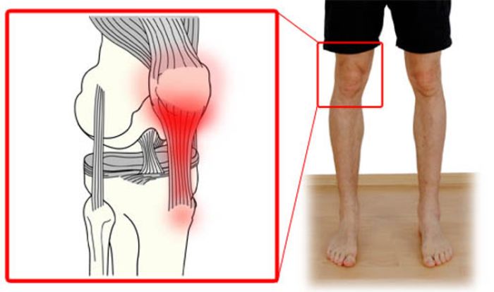 Tendinitis is an inflammatory condition that develops in the patellar tendon, due to twists, tears, or tendon damage. Tendons are fibers that connect muscles to bones. Knee tendinitis occurs most often due to overuse of the knee joint. Patellar tendinitis is known as knee tendinitis. Patellar tendon is an injury that occurs in the tendon that connects the patella to the leg, called a patellar tendon. The patellar tendon works with the muscles at the front of the thigh to extend the knee so you can run, kick and jump. Knee tendinitis is a common condition in athletes whose sports involve running and jumping. But it is not a unique condition of athletes, people who do not perform deportation can suffer from knee tendinitis. Knee tendinitis can be a serious condition that needs attention and treatment, as they can eventually worsen tendon damage and require surgery for treatment, says the orthopaedic in Delhi. Knee Anatomy The knee is composed of 3 bones. The thigh bone that is the femur, the largest leg bone that is the tibia and patella that slides into a groove at the end of the femur. Tendons are strong tissues that connect muscles to the bone. Its size and shape vary, there are small tendons in the fingers and large in the legs. The patellar tendon plays an important role in maintaining the label in place and in straightening the knee. Symptoms of knee tendinitis Pain is the first symptom of knee tendinitis. Pain can manifest directly on the patellar tendon and manifest in cases such as;
In addition, pain may be stiff in the knee, especially when jumping, crouching, sitting, kneeling. Should I see a specialist? Sometimes knee pain can improve with self-care measures and over-the-counter anti-inflammatory drugs, but you may need to visit orthopaedic clinic in Delhi if:
Why does knee tendinitis occur? Knee tendinitis or patellar tendinitis is a common overuse injury caused by constant, repetitive stress on the patellar tendon. Stress causes small tears in the tendon, which as they multiply cause pain and inflammation, explains the sports injury specialist in west Delhi. Risk factors Factors that may contribute to the development of knee tendinitis include:
Complications If no care is received and despite the pain continues to perform activities, it can attract ever larger tears in the patellar tendon. Pain and reduced knee function can persist, if the problem is not addressed, and more serious patellar tendencies can progress, warns the orthopaedic surgeon in Delhi. Get ready for your appointment If the pain persists during or after certain activities and does not improve with self-care measures. Your orthopaedic doctor in Delhi may refer you to a sports medicine specialist. Before you go to your appointment with your orthopaedic in Delhi, you can prepare with:
Diagnosis Knee tendinitis can be diagnosed by reviewing your clinical history, physical knee exam, and performing imaging tests. During the physical exam, your doctor may apply pressure to your knee to determine where you feel pain and may ask you to perform certain movements against resistance. Knee tendinitis pain is usually felt in the front of the knee under the patella. Image testing Some tests that may be suggested to you include:
Knee tendinitis treatment It is usually started with non-surgical treatment. There are several treatment options that can help reduce pain and inflammation. Small tears can be treated with non-surgical treatment. A splint, or brace may be required to provide rest to the tendon and to heal the tendon. But treatment will depend on different factors such as age, level of physical activity, size and type of tendon tear. Therapy Physical therapy can help restore function, decrease pain and prevent future injuries.
Other treatments If nonsurgical treatments don’t help, your orthopaedic in Delhi may suggest other therapies such as:
Prevention To reduce your risk of developing or worsening knee tendinitis, you may:
Recovery Full recovery can take half a year to a year, depending on the extent of the injury and the treatment required.  If you have repeat (inguinal) hip pain and are an athlete or frequently engage in physical activity with discomfort during or after this activity, you may have a hip pinch or hip impingement. A lesion or incipient cartilage can give the first symptoms, explains the orthopaedic in Delhi. The problem is in the case of those who are sedentary, because it is a disease that can appear silently. It has no symptoms, as it does not generate stress on the hip. Here the pain can appear late when the cartilage of the joint is totally worn, causing osteoarthritis of the hip. This disease was discovered 20 years ago. Before its existence, most hip ailments were diagnosed as mono arthritis, causeless inflammation, idiopathic osteoarthritis, unknown cause, or joint wear and tear in late cases. Over time, it was discovered that about 80% of these patients had an alteration in relation to the shape of the bones of the acetabulum with the femur. They already called this hip pinching. This disease contains a lot of different pathologies, it is a set of alterations in the joint, which can be the deformity of the acetabulum or the femur. Symptoms The symptoms of hip impingement can be pain in the groin, discomfort in the hip area around a pain type C, after exercise or appears daily without physical activity. Patients who have been with the disease for a long time and have never had discomfort, the main sign is loss of mobility in the hip. Also, knee pain that you don’t have any findings may be from hip problems. “The knee cries for the hip,” says orthopaedic doctor in Delhi. Symptoms depend on the deformity and physical activity. If there is a lot of deformity and a sedentary lifestyle, symptoms may start. In as much, if there is little anatomical alteration, but the patient is very active, it is more probable that the hip pinch is manifested. And finally, those who have minimal deformity and do little physical activity, it is likely that they can reach the age of 65 and their hip will never hurt. Exams and Diagnosis In the first instance, the orthopaedic doctor in Delhi performs a clinical examination, accompanied by the patient’s symptoms, in which he will move the patient’s leg and, specifically, the hip joint to assess pain and range of motion. To make an accurate diagnosis, the traumatologist indicates a plain radiograph of the pelvis and hip, and an MRI or Artroresonance study. With the radiologist’s evaluation, it is possible to determine where the lesion is, what its size is, if there is a ruptured labrum, tendon injuries, among other types of specifications. What is the Labrum? The labrum is the ring of cartilage, which is located on the outside of the hip joint. It works as a “glue” between the femur and the acetabulum. A labrum tear can occur from high-impact sports or physical activity. Also due to hip abnormalities, says the orthopaedic in Delhi. Treatment The treatment for hip pinching in patients with little deformity, elderly and do not have a significant burden of physical activity, is palliative. That is, it seeks to reduce symptoms, before opting for hip surgery in Delhi, with anti-inflammatory drugs, kinesiotherapy, which has an effect on periarticular inflammation. The other intermediate option is to inject the hip with anti-inflammatory drugs, such as corticosteroids. Which seeks to eliminate acute hip pain. It is a transitory treatment, because the pain will return. If you do little exercise, it will most likely last 1 to 2 years. However, in the case of active people, the hip ailment may go away for 1 month or 1 week. Patients who have a lot of deformity, are athletes and young people, the treating doctor will probably indicate a surgery. Hip arthroscopy Hip arthroscopy in Delhi is a minimally invasive surgery that corrects acetabular and femoral deformities through two holes of 1 centimeter each. Damage is repaired, which is usually the labrum and cartilage. “The surgery lasts around 1 hour and a half. What we do is à la carte”, says the orthopaedic surgeon in Delhi. In addition, in 10% of patients, the muscle area can be intervened, if there is a highly inflamed tendon that has not regenerated, a tenotomy is performed. If there are gluteal problems, the tendon is cleaned. Recovery Once the hip surgery in Delhi is performed, the patient must be hospitalized overnight in the clinic. However, recovery from a hip pinch is slow. During the first 2 or 4 weeks you should use canes. The most important thing for an optimal recovery is the rehabilitation that expert hip kinesiologists must carry out. “People who do poorly rehabilitation can have pain between 6 to 9 months. Those who do a good rehabilitation should not be more than two months with significant pain. Even so, the global rehabilitation to return to play sports, play ball -for example- is six months. If after 6 months I have significant pain, we must start looking for causes of why and treat it, “warns orthopaedic in Delhi. In a year, the patient may have minimal or some periarticular discomfort, but should not have joint pain. 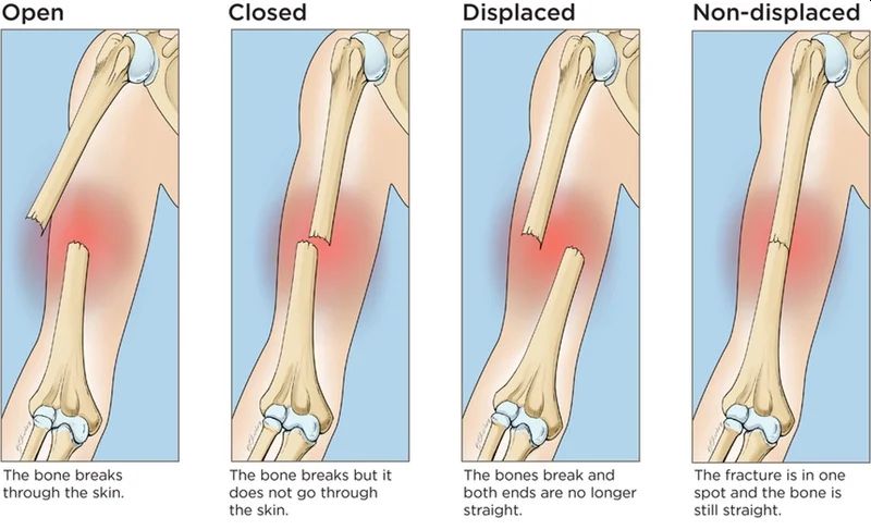 The long bones are part of the skeleton of the limbs. Through them, large-amplitude movements are performed, such as current gestures of daily life (the bones of the thoracic limb) as well as those necessary for walking (the bones of the pelvic limb). The diaphysis of the long bones are frequently exposed to trauma leading to fractures, says the orthopaedic in Delhi. Fractures represent a discontinuity or interruption in a bone as a result of trauma. Humeral shaft fractures are common between the neck and the humeral supracondylar region. Often the location of the fracture is on average 1/3. Humeral shaft fractures are common in adults and much less common in children and the elderly. They can also be found during a difficult labour at birth, with the attempt to release the arm, also called an obstetric fracture. In the case of humeral diaphyseal fractures, the mechanism of production is more frequently indirect compared to the direct mechanism which is much less common. The indirect mechanism determines the fracture with spiroid trajectory (torsion mechanism) or with short and transversal oblique trajectory, in the latter case there is bending by falling on the hand or elbow, explains the orthopaedic in Dwarka. Symptoms Following a fracture, general and local signs are found. The general signs consist of agitation, anxiety, pallor and sometimes the state of shock can be found, especially in important accidents. Impairment of the general condition occurs more frequently in lower limb fractures, in open fractures in both the upper and lower limb, in polytraumas when other visceral injuries occur, explains the orthopaedic surgeon in Delhi. Local signs of fracture may be signs of probability and certainty. The probability signs are:
Signs of certainty confirm the presence of the fracture. These are:
In the conditions of an incomplete fracture (cracks) there are no signs of certainty of the fracture but only signs of probability says the orthopaedic surgeon in Dwarka. Diagnosis and complications Clinical examination and radiography of the humerus establish the diagnosis of certainty. Also, the radiograph at this level can specify if it is a pathological bone fracture. Often humerus is the seat of metastasis to bone spurs visceral neoplasms (breast cancer, lung cancer, kidney cancer, etc.) A complication is immediate radial nerve paralysis, which is impaired in Santa twisting the humerus. The so-called “swan neck hand” appears. Another less common immediate complication is damage to the vessels that irrigate the humerus. The most common late complication is osteoarthritis that develops progressively. A vicious callus is formed which is well tolerated and does not cause functional disorders, only when there is an angle over 20-30 degrees accompanied by a significant gap and a shortening of more than 3 cm. Treatment The treatment is mainly conservative at the orthopaedic clinic in Delhi. It consists in reducing the movements under the action of the weight of the arm and of the external manoeuvres and the immobilization in a plastered thoraco-brachial apparatus with the arm next to the body. If the reduction cannot be achieved, a plastered brachio-antebrachial hanging device can be used, which will favour the reduction in time (continuous traction effect). This device is maintained for 2 maximum 3 weeks and then is replaced with a thoraco-brachial device for another 21 days, says the orthopaedic doctor in Delhi. In the case of fractures that cannot be reduced, with interposition, in transverse fractures, those on pathological bone, surgical treatment is indicated. In oblique fractures, osteosynthesis is recommended by the orthopaedic in Delhi with brooches or rods. In transverse fractures, it is preferable to perform osteosynthesis with the compaction plate. In the case of radial nerve palsy, the fracture is corrected and then the nervous phenomena recede. If no signs of regeneration appear in 6-8 weeks, neurolysis is indicated. Pseudoarthrosis is treated by plaque osteosynthesis and according to the type of pseudoarthrosis by the contribution of spongy autograft. |
AuthorWrite something about yourself. No need to be fancy, just an overview. Archives
August 2022
Categories |
 RSS Feed
