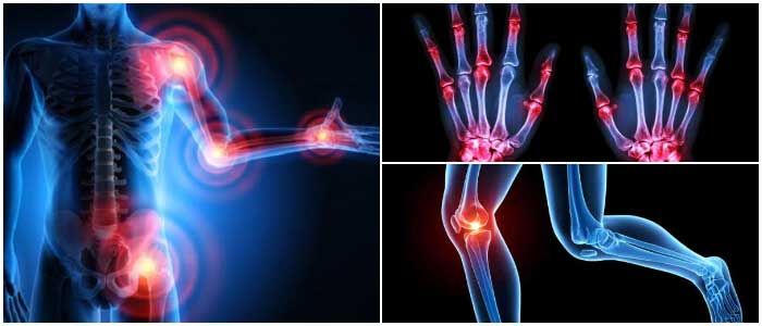
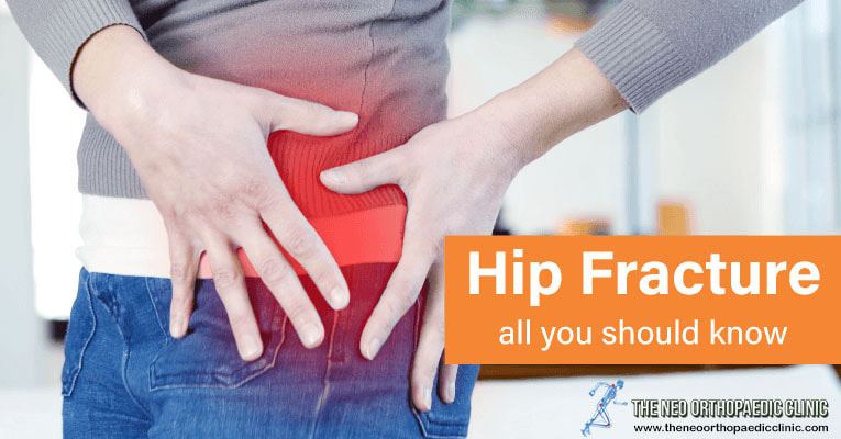
 Overview A hip fracture means more than a broken bone. For the elderly, hip fracture means a major change in life. Most likely, surgery will be needed, and recovery may take more than a year, says orthopaedic surgeon in Delhi. The activity and physical therapy, but also the help of the family and a caregiver will contribute to the recovery of mobility. Most hip fractures are suffered by people over the age of 65. Those in this age group must be very careful to avoid falls. In most people, the hip fractures in the upper thigh (femur), close to where the thigh bone joins the hip joint. Symptoms Signs and symptoms of a hip fracture include:
Causes Falls are the leading cause of hip fracture in older adults. As people age, their bones become less resilient and are naturally prone to breakage, even after minor trauma. Children and young adults are more likely to have hip injuries, says the orthopaedic in Delhi. The causes of hip fractures include:
Risk factors Among the risk factors that can increase the risk of rupture of the hip bones are:
Complications A hip fracture is a serious injury. Although the fracture itself is treatable, complications can endanger a person’s life. If a person suffers from a hip fracture, surgery may be necessary. The orthopaedic in west Delhi may use an external traction system that will allow the hip to heal. The biggest risk when using this system is that it can cause muscle damage and weakness, increasing the likelihood of permanent loss of mobility. In addition, traction will keep the patient immobilized for a long time, during which time blood clots can develop in the veins of the legs. Affected veins can be located on the surface of the skin, causing thrombophlebitis superficial or may be located deeper, in the muscles, resulting in deep vein thrombosis. The risks of using the traction system include:
Treatment Your orthopaedic doctor in Delhi will advise your patient to exercise as soon as possible after surgery. This helps prevent complications such as pneumonia, blood clots and scabies. After the operation, it will be difficult for the patient to do many of the activities alone, so it may be necessary to be admitted to a rehabilitation center for a period after the operation. The more active a person is, the faster he will recover. Ways to prevent There are many steps that can be taken to prevent a hip fracture. One of the most important is to prevent osteoporosis, which can occur in both women and men. To slow down and prevent osteoporosis the orthopaedic in Dwarka advices to follow:
0 Comments
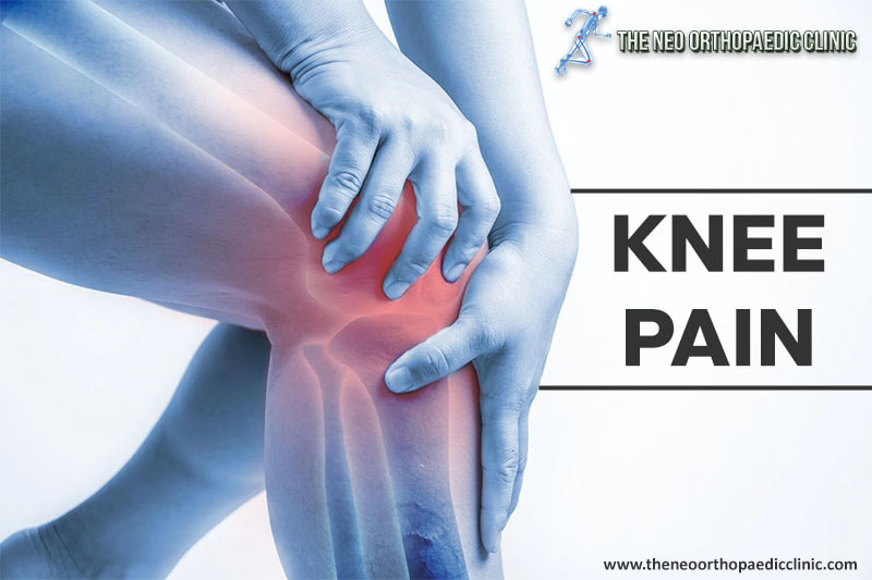 Overview Knee pain is one of the most common ailments among the population. Some of the most common reasons for knee pain are inflamed or torn ligaments and certain cartilage conditions, says orthopaedic surgeon in Delhi. The knee is a complex joint and there can be several factors that cause pain. In order to find out the reason for your knee pain, you will need to perform a thorough examination, such as an MRI or a special type of MRI, in which a dye is injected into the knee so that the joints can be seen in detail. The treatment is prescribed and recommended after establishing the diagnosis, only by the orthopaedic in Delhi. The main ailments Bursitis. The bursa is a fluid sac that acts as a buffer and protects the joints in several areas of the knees. Overuse, falls can irritate the bursa, causing pain and swelling in the knees. Iliotibial tendon syndrome. The iliotibial tendon is a hard tissue that makes the transition from the hip to the lower tibia. If it is irritated due to overload, it may become inflamed and cause pain on the outside of the knee. Osgood-Schlatter disease. This condition leads to a painful inflammation, located below the knee, where the tendon at the patella “connects” to the tibia. It is caused by overload and irritation of the tendon; the pain can appear and disappear without treatment. Osteoarthritis. This is a common cause for knee pain in athletes or non-athletes, but also for those over 60 years of age. Tendonitis or swelling of the tendons. Tendons are strips of tissue that connect bones and muscles, and overuse leads to their inflammation. If not treated properly it can also cause knee pain. Gonarthrosis. It is a degenerative condition of the knee joint, which arises from processes of degradation of joint components, especially the articular cartilage, as well as due to greater efforts than the knee joint can withstand. The main causes of gonarthrosis are: standing for a long time, walking for a long time, carrying weights, but also problems related to overweight, as well as certain injuries. Unfortunately, over time, this disease progresses quite a lot, especially in the absence of specialized treatment by orthopaedic in Dwarka. At first, the patient hears sounds like cracks in the knee joint, then there is pain for a long time standing. When the disease is already in a more advanced stage, the pain appears more and more often while walking. There is basically a limitation of knee mobility. In the final stage of the disease, the patient will experience persistent pain including rest, and the knee joint becomes swollen and increases in volume. How to prevent knee pain
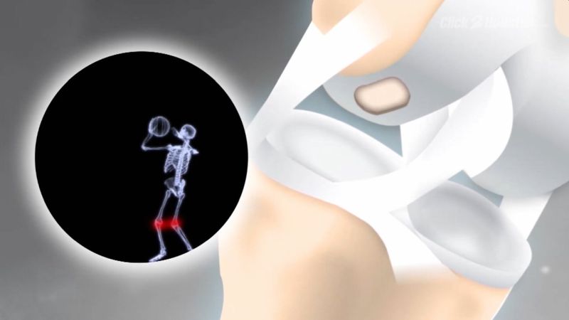 WHAT IS THE CARTILAGE IN THE KNEE FOR? An approx. 5 mm thick layer of cartilage covers the bone in the knee joint. The smooth surface of the cartilage allows the joint to move smoothly. In addition, the cartilage causes an even distribution of pressure and shock loads on the underlying bones. This protects the bone and prevents or reduces overstressing. The cartilage has no nerves and stops growing after puberty. This layer of cartilage accompanies us throughout life. HOW DOES CARTILAGE DAMAGE COME? The cartilage undergoes natural aging. In doing so, it loses the ability to store water and the cartilage layer shrinks. The surface of the cartilage becomes brittle and cracks. This brittle surface is more prone to impacts and shear forces. This allows the cartilage to wear out or split off more quickly. Since the cartilage has no pain fibers, we do not feel this change at the beginning. Perhaps a rubbing noise (crepitation) is noticeable under greater stress. In contrast to cartilage, bone has pain fibers. If the bone is exposed, we feel the affected joint with the corresponding pain, explains the orthopaedic in Delhi. However, the articular cartilage can also be damaged by chronic stress or an accident (trauma). HOW DO YOU RECOGNIZE CARTILAGE DAMAGE? With acute cartilage damage, patients complain of blockage of the knee joint, swelling and pain. The extent of the discomfort depends on the size and depth of the cartilage defect and its location. In the case of chronic cartilage damage, patients report start-up, stress, and inflammatory pain. The knee is swollen, and the mobility of the knee joint is limited. In addition, the patients have an unsteady gait, the knee joint feels unstable and in some cases kinks, says the orthopaedic in Delhi. HOW IS CARTILAGE DAMAGE DIAGNOSED? The damage to the articular cartilage can appear superficially with small cracks on the one hand, but on the other hand affect the entire cartilage in the knee. This causes the rough, painful surface of the bone to emerge. Cartilage damage is divided into four stages:
In addition to the exact questioning (anamnesis) and well-founded clinical examinations of the knee joint, X-ray and MRI images are necessary. Based on these documents, the appropriate individual therapy can be discussed, says the orthopaedic doctor in Delhi. WHAT TREATMENTS ARE AVAILABLE FOR CARTILAGE DAMAGE? The cartilage damage looks different depending on the cause, whether accidental (acute) or wear-related (chronic) and is treated differently accordingly. In the case of acute cartilage damage, for example, we have a clearly defined defect (punch defect) compared to healthy cartilage with sharp edges. This is not the case with chronic cartilage defects. If the cartilage defect is not treated, there is further cartilage wear of the knee joint and, in the further course, knee joint osteoarthritis. Conservative therapy for cartilage damage is very limited. After puberty, the cartilage loses its self-healing potential, i.e. from this point onwards we have to get by with the cartilage for our entire life. For these reasons, the natural course of cartilage damage leads to deterioration. The cartilage damage gets bigger and deeper over time, which leads to a clinical deterioration with corresponding pain and restrictions in everyday life and during sporting activity. With conservative therapy, cartilage damage cannot be cured, but only alleviated by slowing down the wear and tear of the cartilage. The following conservative therapies are possible:
The surgical therapy of the cartilage damage depends on the size (extent) and depth of the defect and must be individually adapted. In addition, factors such as the integrity of the exposed bone, cartilage quality on the opposite side of the defect and the age of the cartilage defect play a decisive role, explains the orthopaedic surgeon in Delhi. Furthermore, the younger the patient, the greater the chances of success of the methods described below for acute cartilage damage. These include:
The above-mentioned surgical techniques are mainly used for acute cartilage defects. In addition to age, the opposite side of the cartilage defect also plays a role. This should not show any major damage, otherwise the rough surface can negatively affect the outcome of the operation. Another option for covering more chronic cartilage defects is knee replacement in Delhi of the injured cartilage. If the cartilage wear has progressed so far that finally bone rubs on bone (end stage of osteoarthritis), a partial prosthesis (e.g. a sled prosthesis, a kneecap glider replacement) or a knee replacement surgery in Delhi is necessary. A correction of bowlegs / knock knees (so-called corrective osteotomy) is sometimes necessary. WHAT IS THE AFTER-TREATMENT LIKE? The follow-up treatment must be individually adapted to the previous operation. If cartilage therapy was carried out, the knee joint had to be relieved with walking sticks for six weeks. In the case of cartilage defects behind the kneecap, mobility is also restricted for six weeks. Patients with resurfacing can immediately put weight on and move the knee joint. To protect the soft tissues, however, walking sticks are recommended for around four weeks. Physiotherapy for several weeks to relieve swelling and strengthening and stretching of the thigh muscles are also important. 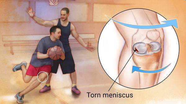 Very often we hear people (young and old) with knee discomfort or accidents, which is one of the most common injuries, and end up sometimes undergoing meniscus surgery. With this article orthopaedic in Delhi explains you what meniscus are and how to treat your injury. Definition of meniscus injury: Meniscuses are fibrocar cartilage that are located inside the knee joint and are located between the femur and tibia. There is a medial meniscus (inner part of the knee) and another side (outer part). It has a semi-lunar shape and its main function is to increase the depth of the relatively flat surface of the top of the tibia and be able to be a true cushion of the knee. In younger patients, the meniscus is a fairly resistant and elastic structure and the rupture of the meniscus is caused by a significant twisting or turning of the knee. In older people (40–45 years) who perform a sport, the meniscus becomes weaker, the tissue degenerates and is less resistant and the injury can be caused by minor trauma, for example, by rising from the squat position or by performing an exaggerated bending of the joint. Usually, this type of injury is very much presented in contact sports such as football and rugby and is suffered by athletes in general. 1. Symptoms of meniscus injury: The most common symptoms of meniscus tearing are:
2. Self-care to prevent meniscus injury: It is always advisable to have a proper physical preparation, but not only from an aerobic point of view, but also from the point of view of strength and toning, which is the sense that alerts the body to the location of the muscles. Injuries are more common in people who do occasional physical activity and with very low physical preparation, says orthopaedic doctor in Delhi. 3. Treatment of meniscus injury: It should always be evaluated by a specialist doctor and your orthopaedic surgeon in Delhi will treat your tear depending on the type of tear, size and location. If the injury is longer and surgery has had to be performed, the period of return to normal activity will be longer and under specialized medical supervision. In addition, depending on the type of tear you have, your age, activity level, and any related injuries, they will be factors that will influence your treatment plan. 4. Non-surgical treatment in meniscus: If your tear is small and located on the outer edge of the meniscus, it may not require surgical repair. As long as your symptoms don't persist and your knee is stable, nonsurgical treatment might be all you need. THE RICE protocol for meniscus injuries This protocol is effective for most sports-related injuries. RICE stands for R-Rest, Ice, Compression, and E-Elevation.
|
AuthorWrite something about yourself. No need to be fancy, just an overview. Archives
August 2022
Categories |
 RSS Feed
